KINSC Imaging Contest Winners Announced

Audra Devoto '17 (First Place Winner)
Details
First Place: Audra Devoto '17
Second Place: Rui Fang '17
Third Place: Matteo Miazzo '15
The KINSC Scientific Imaging Contest is an annual contest for student-submitted images from experiments or simulations that are scientifically intriguing as well as aesthetically pleasing. Judging is based on both the quality of the image and the explanation of the underlying science. First, second, and third place winners will have their images displayed on the walls of the KINSC.
Audra Devoto '17 (First Place Winner)

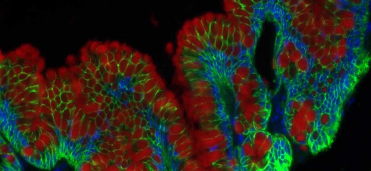
The effect of Interleukin 13 (IL13) on cultured small intestine epithelial cells from a normal patient. The cultured cells show several crypts and villi characteristic of the intestinal epithelium. Cell membranes are stained green, mucin is stained red, and cell nuclei are stained blue. IL13 is a cytokine involved in inflammatory and anti-inflammatory pathways in the airways and intestinal epithelium (1). Here, the response of healthy small intestine epithelial cells to IL13 treatment is shown to involve goblet cell (mucin-producing cells) hyperplasia and mucin hypersecretion.
Rui Fang '17 (Second Place Winner)

A plastic fork viewed through crossed polarizers shows beautiful multicolored patterns. This is due to birefrigence of plastics. In birefringent materials, the index of refraction of a plane-polarized light can take on two values, resulting in a phase retardation which changes the polarization of transmitted light. Different colors of light have different indices. So, through another polarizer, a fringe pattern is revealed due to color interference. Because the pattern is indicative of the varying birefringence of the material, this method called photoelasticity is often used for analyzing stress distribution.
Matteo Miazzo '15 (Third Place Winner)

As part of our research, we induce controlled flow using electric and magnetic fields to produce Lorentz forces within a conductive fluid. In order to characterize this flow field, we need a way to visualize the motion. Tracer particles are seeded on top of the fluid; these particles are tracked and the paths are analyzed, allowing us to determine specific characteristics of the flow field. This image shows the paths of individual tracers, which have been randomly colored to distinguish between different particles.


