2023 KINSC Scientific Imaging Contest Winners Announced

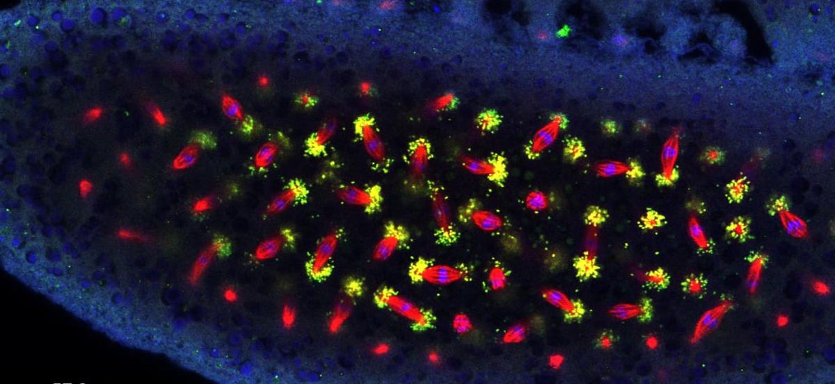
The winning image, by Ash DiCristofalo '23, of this year's 2023 Scientific Imaging Contest.
Details
The KINSC Scientific Imaging Contest is an annual contest for student-submitted images from experiments or simulations that are scientifically intriguing as well as aesthetically pleasing.
Judging is based on both the quality of the image and the explanation of the underlying science.
First Prize: Ash DiCristofalo '23

"Bacteria Disco" shows the intracellular bacteria Wolbachia (green dots) localizing along microtubules (red lines) during cell division (with blue DNA dots at the microtubules' centers) in a Drosophila sturtevanti (fruit fly) embryo. My senior thesis used antibody staining and confocal microscopy of three species of Drosophila fruit flies to test the hypothesis that the localization of Wolbachia along cells’ astral microtubules during fly embryonic development is conserved across species. In this image, the cells within this embryo are dividing, and the Wolbachia is present at the ends of the microtubules as the spindle lines up the chromosomes for division.
Second Prize: Annemarie Wood '23

While marine pollution is often in the news for its negative environmental impacts, plastic debris also provides an alternative habitat for ocean organisms. Animals, plants, and bacteria accumulate on plastic surfaces, forming little communities that can be carried by ocean currents far from their original territory. Hitching a ride on the plastic bottle shown here are several barnacles and a calcareous casing that formerly housed a marine tubeworm. By covering the surface of the plastic, these organisms may play a protective role in preventing its breakdown via photooxidation from the sun.
Third Prize: Darshan Patel '23

The Triangulum Galaxy is the third largest galaxy in our Local Group, providing an ideal opportunity to investigate the dynamics of spiral galaxies due to its proximity and orientation. Its abundance of HII regions (in red), which act as nurseries for star formation is noteworthy, with NGC 604 being the brightest and housing over 200 young O-type stars. The data used to create this image was collected on October 14, 2022 using the 16” telescope at Strawbridge Observatory for the “Advanced Topics: Observational Astronomy” course. The data was later processed using AstroImageJ and Photoshop to highlight the galaxy's unique features.
Honorable Mention: Charlie Mamlin '23

Scanning electron microscopy (SEM) image of ~300 million year old Calamites water transport cells (tracheids). Calamites is an extinct genus of land plant found in swamp ecosystems during the Carboniferous Period: they looked like—and are closely related to—modern horsetails, but were the size of trees. After being dissolved in acid and gold-coated, Calamities fossils can be examined under SEM to measure the dimensions of the porous spaces (pits) in their tracheids. Using these measurements, we model water flow through these extinct plants to further investigate the evolution of plant hydraulics and how plants responded to past episodes of climate change.
Honorable Mention: John Dvorak '23

This is a Z-stack projection of a seven-day-old transgenic zebrafish larva induced with 4-hydroxytamoxifen (4-OHT) at 28 hours. The fish was mounted on its side in low-melting point agarose and imaged under our new confocal microscope. Here, all cells are programmed to express a green fluorescent protein, while in some cells 4-OHT has induced a recombination event that leads to red expression. The resultant green-to-red switching is visually striking as individual cells, including long muscle cells, are accentuated.
Honorable Mention: August Muller '23

The Eagle Nebula (M16) is an astronomical object in the Serpens constellation, famously known for its ‘Pillars of Creation’ feature. Only about 7,000 lightyears away, M16 is quite large on the sky (similar to the size of the Moon). Its recognizable star-forming regions have been imaged here using an H-alpha emission line filter (magenta), along with O-III emission (cyan) and clear (yellow) filters. The abundance of star-forming H-alpha is clear from the purple color of the nebula in the image, though emission in the other filters is present. Image taken using Haverford College’s Strawbridge Observatory 16” telescope.
Honorable Mention: Jahsaiah Moses '23

Wolbachia utilize microtubules for transport in the cell. The relationship has allowed for Wolbachia to develop a weak competitor status as co-opting cellular structure would prove disadvantages to the vertical transmission patterns. Wolbachia have succeeded in manipulating the reproduction of their host and increasing the bacterial spread in insects. Embryos were fixed with methane/heptane fixation protocol then immunohistochemistry staining to isolate DNA, Wolbachia and tubulin with Anti-Hsp60 primary antibody and secondary antibodies including Hoechst DNA stain (blue), Alexa Fluor488 (green) and Alexa Fluor 546 (red), respectively.


