2017 KINSC Scientific Imaging Contest Winners Announced

Details
The KINSC Scientific Imaging Contest is an annual contest for student-submitted images from experiments or simulations that are scientifically intriguing as well as aesthetically pleasing.
Judging is based on both the quality of the image and the explanation of the underlying science. First, second, and third place winners will have their images displayed on the walls of the KINSC.
First Place: Annika Salzberg '19

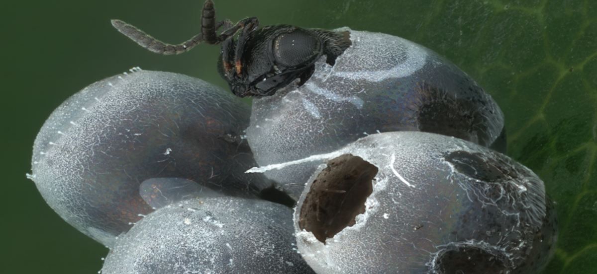
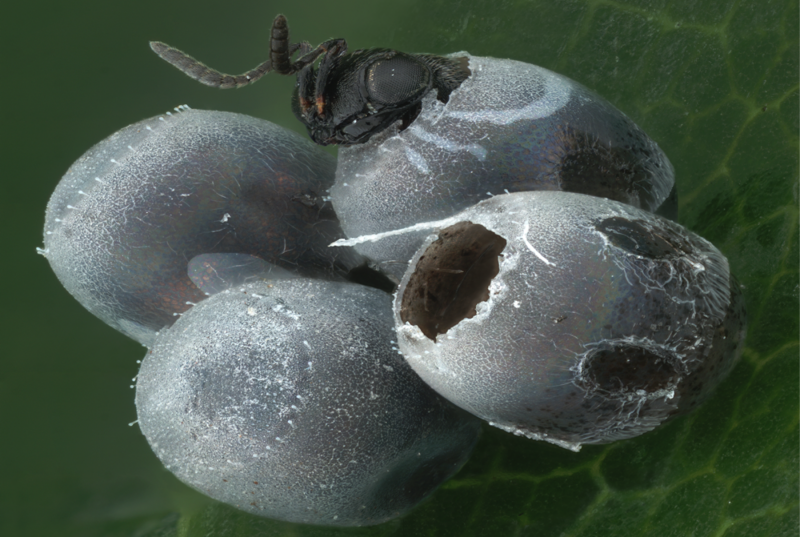
Trissolcus japonicus, a parasitic wasp in the superfamily Platygastroidea, emerges from a hemipteran egg. This wasp and other parasitoids of hemipterans are being studied for their possible use as a biological control agent against invasive brown marmorated stink bugs. This image was created at the Smithsonian Museum of Natural History under the supervision of Dr. Elijah Talamas using stacked light microscopy and Adobe Photoshop. It consists of over 42 stacked images, each containing between 15 and 80 images each. Special credit: USDA/Systematic Agricultural Research Service
Second Place: Kate Rose Sullivan '18

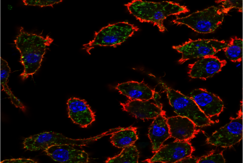
Macrophages release calreticulin, an endoplasmic reticulum chaperone protein, to bind to target cells. Calreticulin binds to unique glycoepitopes on the cell surface. The regulation of this glycoepitope mediates the cellular localization of calreticulin and, ultimately, immune recognition for tissue homeostasis. This image demonstrates the cellular distribution of the glycoepitopes (green) within macrophages that are actively releasing calreticulin. Cell surface glycoepitopes are observed outside of phalloidin stain (red), which labels actin filaments at the periphery of the cell and dapi (blue) staining the nucleus. Confocal images were acquired in the laboratory of Dr. Irving Weissman at Stanford University and was funded by the KINSC through the Frances Velay Summer Scholars Program.
Third Place: Gabe Oppler '17

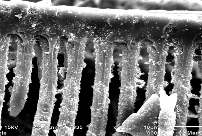
Detailed view of scalariform pitting on a stem xylem tracheid of Psaronius, an extinct tree fern from the Pennsylvanian subperiod of the Carboniferous (323-299 Ma). This tracheid was embedded in a fossil from the West Mineral, KS formation. The fossil was macerated in hydrochloric acid to liberate the organic material as part of Gabe Oppler’s investigation into the stem and root anatomy of Psaronius. Imaged under the Haverford College Scanning Electron Microscope (SEM) at 2,200x magnification.


