2019 KINSC Scientific Imaging Contest Winners Announced
Details
The KINSC Scientific Imaging Contest is an annual contest for student-submitted images from experiments or simulations that are scientifically intriguing as well as aesthetically pleasing.
Judging is based on both the quality of the image and the explanation of the underlying science. First, second, and third place winners will have their images displayed on the walls of the KINSC.
First Place: Daniel Feshbach '20
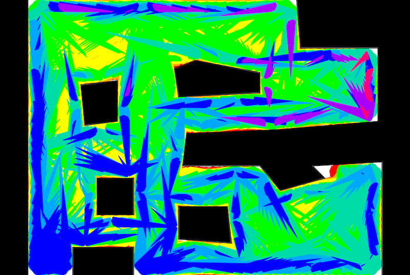
In robotics, coverage considers how to pass over an environment. It is useful in cleaning, mine removal, and more. Here we plan paths for a circular robot which moves at an unreliable angle until encountering a wall. We model the uncertainty and assume worst cases to plan paths which guarantee to cover the depicted regions. Environment walls are black, and colors represent the coverage guarantee under upper bounds on the direction error. They are stacked in increasing order, so the red area (≤ 0.5 ◦ error), while largely obscured, is the largest coverage guarantee because it has the most precise movements. —Daniel Feshbach '20
Second Place: Reilly Milburn '19

On a KINSC funded trip to attend the International Pulsar Timing Array annual conference in New Mexico, we were given a tour of the Very Large Array. The VLA consists of 27 25 meter diameter radio antennas positioned in a Y configuration. The size of the configuration can be changed depending on the type of radio observing taking place. Radio telescopes allow us to see objects we normally wouldn’t see in optical wavelengths. With the use of radio telescopes such as the VLA, Professor Andrea Lommen’s collaboration is able to time pulsars in hopes of detecting gravitational waves. —Reilly Milburn '19
Third Place: Renata DiDonato '19
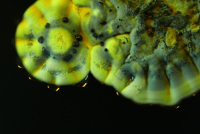
Streptomyces coelicolor is a Gram-positive bacteria belonging to the phylum actinobacteria. This soil-dwelling microorganism biosynthesizes the natural product actinorhodin, a blue-pigmented polyketide antibiotic. When actinorhodin is released into the strain’s growth medium, the medium turns a deep blue. A Streptomyces coelicolor spore grown on a semi-solid agar plate is depicted here. In this image the bacteria has initiated its chemical warfare, releasing actinorhodin into its environment. —Renata DiDonato '19

