Strengthening Science

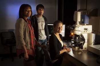
(left to right) Shanaye Jeffers, Faraz Sohail and Kaitlyn Shank, all '12, use the new confocal microscope to do research on the progress of neurodegenerative diseases in nematodes for their Bio300 class, also known as Super Lab.
Details
In the fall of 2009, Haverford's departments of biology and physics were awarded an exceptional $1 million grant from the National Science Foundation (NSF) to purchase four high-tech imaging instruments. One year later, all of the instruments have arrived on campus, and students and faculty are reaping the benefits of enhanced capabilities for their teaching and research programs.
First to arrive in October of '09 was the fluorescence-activated cell sorting system (FACS), which can analyze various features of individual cells and then sort or extract cells with specific characteristics. Last year, students in the junior“Superlab” used the FACS in an experiment designed by post-doctoral fellow and instructor Bob Daber. While examining the evolution of a sugar binding site in a bacterial system, the FACS allowed students to sort bacteria that glow green when related sugars activated a certain gene.
Haverford seniors have taken charge of operating the FACS, says Professor of Biology Jenni Punt.“I believe they are the only undergraduates in the country to run such a machine,” she says.
Also arriving on campus last semester was a confocal microscope. Unlike a regular microscope, which can focus on the forefront of an image while leaving the background blurry, the confocal microscope slices layers from three-dimensional objects like cells, tissues and organisms and brings those layers into sharp focus. Professor of Biology Phil Meneely's students have used the microscope to study specific changes in chromosomes during meiosis, a process in which the number of chromosomes per cell is cut in half.
“The students have been 100 percent responsible for this instrument,” says Meneely.“They trained themselves on it and have even written a users' manual.”
A scanning electron microscope, which arrived in March, is used in labs overseen by Assistant Professor of Biology Rachel Hoang and Professor of Physics Walter Smith. The instrument provides Hoang's students with detailed views of the surface structures of cells, which has contributed to their research on the evolution of genetic pathways. Students in Hoang's lab have also used a College“Teaching with Technology Grant” to develop lab manuals, video instruction segments and an integrated website for all of the instruments.
The final instrument to arrive was the transmission electron microscope. It offers molecular-level detail of atoms, proteins, and cells.“It's wonderful to be able to walk through a cell as you're sitting at the microscope,” says Karl Johnson, Professor of Biology and department chair. Johnson co-authored the original NSF grant proposal with Hoang, Punt, Smith, and Professor of Biology Rob Fairman.
Haverford has also hired an instrument specialist, George Neusch, who will help maintain the equipment as well as train and serve as a resource for students. This position was funded through support from alumni and private foundations.
-Brenna McBride


